2015-12-09
Horse Eye Injuries and Ailments
Posted at 20:15h
in Uncategorized
by Arizona Equine | Arizona Equine Medical & Surgical Centre
0 Comments
Did you know that the horse has one of the largest eyes of any land mammal; the average size of a horse’s eye is approximately 34mm in diameter. Of the land mammals, the moose passes the horse up with an eye approximately 40mm. The largest mammal eye is the blue whale measuring in at 150mm! Of all animals, invertebrate and vertebrate alike, the all-time winner is the Colossal Squid with an eyeball measurement of 279 mm.
Horse Eye Injury
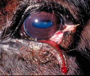
Horse eye injuries occur from a variety of causes ranging from getting poked by hay, rubbing their eye, to eyelid lacerations and skull fractures. A brief review of some of the common injuries includes ulcers, foreign body injury, lacerations, orbital fractures, uveitis, and tumors.
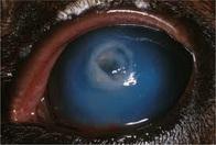
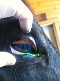
- Ulcers – this injury is a scratch or abrasion to the cornea (the clear portion of the eye). A corneal ulcer is very painful, owners will see tearing, squinting, and the horse will squeeze their eyelids together. It is imperative that your veterinarian examine your horse as soon as possible. Your horse may need to be sedated, block the eyelid (nerve block to allow the veterinarian to open the eyelid), and stain the eye to determine the extent of the ulcer. A small amount of fluorescein stain (a green dye) is applied topically to the eye. This stain will adhere to exposed connective tissue in the cornea, and allows us to fully see the size and depth of the ulcer. If necessary, some debridement may be performed to remove any loose edges of the ulcer prior to initiating treatment. If severe trauma occurs, or a simple ulcer is not tended to, a more complex ulcer can form. These ulcers tend to affect more layers of the cornea. One type of ulcer is called a “descemetocele”. (Deh-seh-met-o-seel) (Descemet’s membrane is the second to last layer of the cornea). These tend to appear as white opacities with a black spot in the middle. That black spot means that there is only one cell layer left between the outside world and the inside of the eye. If we were to stain this eye, we would see a rim of green dye with a black hole in the middle.
 Ulcers such as this put the eye in grave danger of rupturing and need to be treated more aggressive therapies, sometimes needing hospitalization. Treatment usually consists of ophthalmic antibiotic ointment, atropine ointment (a dilating medication to relax the iris which can spasm), and a non-steroidal oral medication such as Phenylbutazolidin (Bute) or flunixin meglamine (Banamine). DO NOT APPLY OPHTHALMIC OINTMENT WITH STERIOD AS THIS WILL IMPAIR HEALING.
 - Foreign bodies – are items that include but not limited to hay, wood, and burrs. These items can be imbedded in the eyeball, conjunctiva or simply floating onto of the eye. If the foreign body is not noticed it can result in an ulcer. You may notice increased tearing, squinting, and actually see the foreign body. The veterinarian commonly sedates the patient, blocks the eyelid, and removes the offending article. However, if the foreign body is imbedded in the eyeball, your horse may require surgery to remove the agent. Treatment commonly includes antibiotic ophthalmic ointment, oral systemic anti-inflammatory medication, and possibly oral antibiotics.
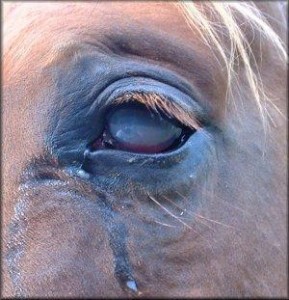
- Eyelid lacerations – the upper eyelid is critical in maintaining lubrication to the eye. These injuries commonly occur when the horse rubs their eye on a bucket, fence or a sharp object in the stall. Even if you suspect the laceration is greater than 8 hours old, you should still contact your veterinarian. Head and eyelid lacerations have quite a bit of blood flow and therefore once the wound is cleaned up a surgical repair can be performed. Surgical repair requires precise alignment of the damaged tissue to prevent healing tissue from rubbing on the eyeball and creating an ulcer. The horse often wears an “eye saver” to prevent them from rubbing their eye and hence rubbing the sutures out. Again, ophthalmic antibiotic ointment, systemic (oral) antibiotics and non-steroidal anti-inflammatories are usually recommended.
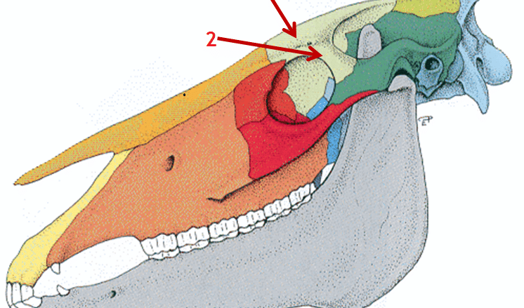
- Facial and orbital fractures – the horse owner usually sees a swelling, a depression, and sometimes a laceration associated with the fracture. Most fractures require radiographs (x-rays) to determine the extent of the damage. If there is a non-displaced fracture surgery may not be required. However, a complex and/or displaced fracture will be required to maintain the orbit and provide protection to the eyeball.
- Uveitis – can be an acute or chronic problem. Uveitis occurs when there is inflammation within the eyeball, the inflammation can be due to and infection or a non-specific immune response. This is a very painful condition and the horse will squint, squeeze their eyelids closed, and have tearing. The veterinarian will differentiate uveitis from the similar clinical signs of a corneal ulcer. With uveitis, the cornea does not necessarily “take up” the fluorescein stain as it does with an ulcer. Treatment utilizes ophthalmic ointment and possibly systemic steroids (such as prednisolone).
- Tumors – common tumors are eyelid sarcoids and conjunctival squamous cell carcinoma. Sarcoids are one of the following: occult, verrucous, nodular, fibroblastic, mixed or malevolent. Treatment depends on the type of sarcoid. Squamous cell carcinoma is best treated early as this type of cancer grows rapidly. Squamous cell carcinoma is commonly located on the horse’s third eyelid. Removal of the affected portion of the third eyelid is usually the treatment of choice.The key take away concept with horse eye injuries and ailments is to contact your veterinarian as soon as you suspect a problem. Rapid treatment will help ensure maintaining your horse’s vision.

Sorry, the comment form is closed at this time.